

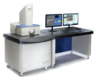
TEM sample preparation system picomill M1080
Description
Combines an ultra-low energy, inert gas ion source, and a scanning electron column with multiple detectors to yield optimal TEM specimens.
Key Specifications
- Achieve ultimate specimen quality – free from amorphous and implanted layers
- Complements FIB technology
- Milling without introduction of artifacts
- Advanced detector technology for imaging and precise endpoint detection
- In situ imaging with ions and electrons
- Microscope connectivity for risk-free specimen handling
- Adds capability and capacity
- Fast, reliable, and easy to use
– Yield enhancement
– Failure analysis
Specification
Applications
Primary: Microelectronics and semiconductor transmission electron microcopy (TEM) specimen preparation
Secondary: Any other specimens requiring optimal results Ideal for when FIB preparation is combined with aberration corrected TEM
Ion source
Filament-based ion source combined with electrostatic lens system Variable voltage (50 eV to 2 kV), continuously adjustable Beam current density up to 8 mA/cm2 Beam size < 1 µm
Electron source
Accelerating voltage up to 10 keV Working distance of 16 mm Faraday cup for electron beam current monitoring with a range of 1 to 2,000 pA
Goniometer
TEM style X, Y, and Z axes movement and α tilt Specimen exchange of < 30 seconds Milling angle range of −15 to +90° Viewing range -15 to 180°
Holder
Side-entry, TEM-style holder Compatible with all major TEMs
Ion beam targeting
Ion beam can be targeted to a specific point on the specimen surface or scanned within a selected area
User interface
Menu-driven with system status displayed
Gas Ion source
gas: UHP 99.999% argon
Gas control: Automated using mass flow control technology
Pneumatic supply: Compressed dry air or dry nitrogen at 2 to 7 bar
Imaging
Secondary electron detector/Everhart-Thornley detector
- Electron imaging with 2 mm field of view
- Ion-induced secondary electron imaging with 1.9 mm field of view
- Specimen image displayed on PicoMill system user interface
Solid-state backscatter electron detector
Solid-state scanning/transmission electron (STEM) detector
Vacuum system
Turbomolecular drag pump backed by an oil-free diaphragm pump
Specimen chamber pressure:
- Base vacuum of 3 x 10⁻⁶ mbar
- Operating vacuum of 1 x 10⁻⁴ mbar
Electron column: Base vacuum of 1 x 10⁻⁶ mbar
Specimen goniometer: Atmosphere to 1 mbar (pre-pump)
Vacuum gauges:
- Cold cathode for specimen chamber and electron column
- Pirani gauge for goniometer
Automatic termination
Termination by time, electron signal, or manual process
Accesories
Consumables
https://micro-shop.pl/kategoria-produktu/tem/siatki-z-pokryciem-carbon/
For more supplies, please visit our online store Micro-Shop.