

Phenom Pharos
Description
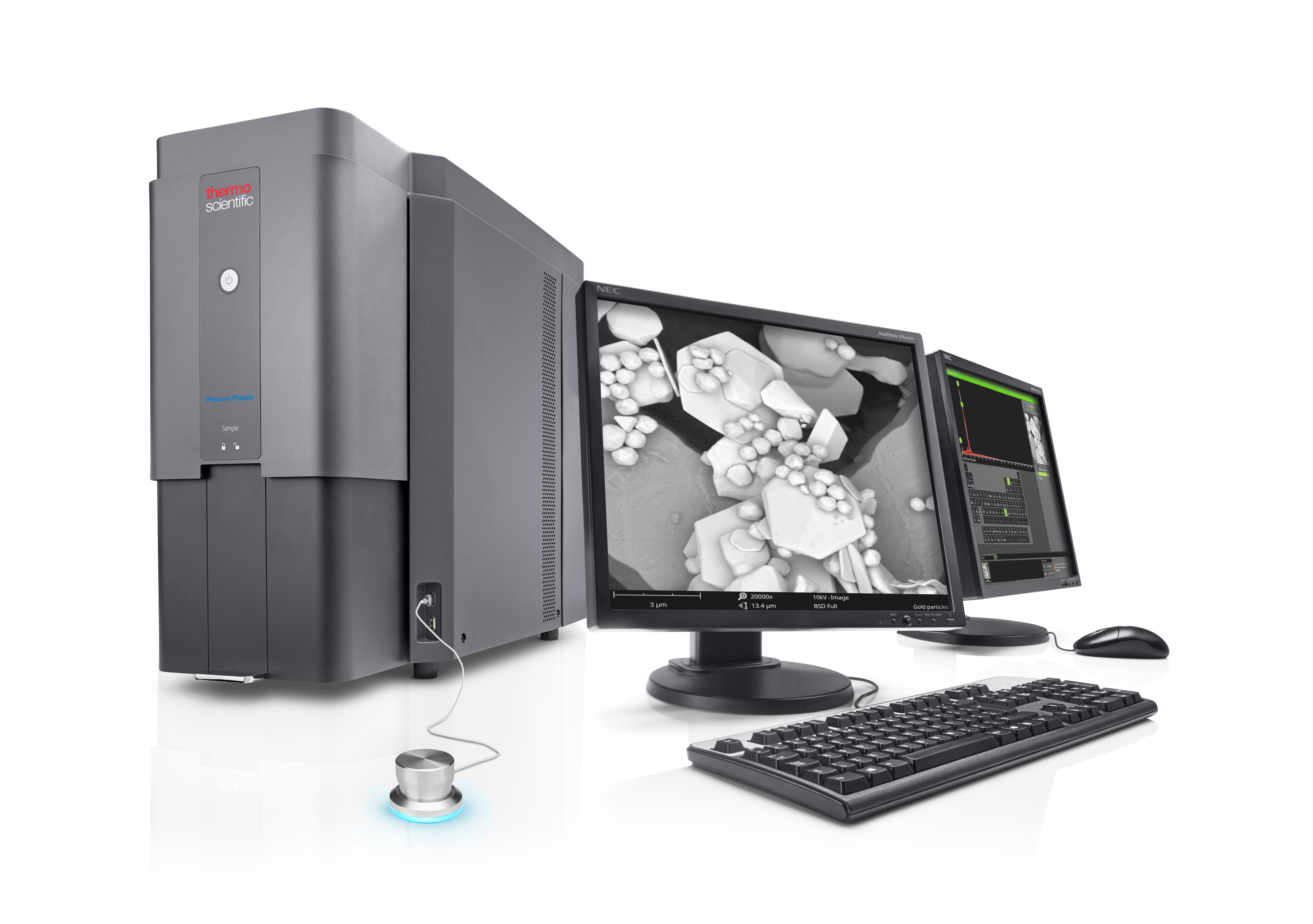
The Phenom Pharos desktop scanning electron microscope (SEM) is brand new model by Thermo Fisher Scientific. Fastest and higher resolution SEM of all Phenom models . With its long-life high-brightness FEG (Field Emission Gun) electron source, the Phenom Pharos creates state-of-the-art images with a minimum of user maintenance intervention.
The Phenom Pharos microscope is dedicated to easly acquire SEM images of highest quality with magnification up to 2 000 000x and image resolution less than 2 nm.
The Phenom Pharos microscope is the fastest SEM in the market. The time from loading the sample up to acquiring SEM image is less than 30 seconds.
A large variety of sample holders are available for facilitating the fast loading of any sample into the Phenom. From long axial-shaped samples to moist biomaterials, there is always a suitable holder for the sample to be analyzed.
Like in case of other models Phenom Pharos can be upgraded by many kinds of sample holders and advanced application software.
The Phenom Pharos is equipped with highly efficient FEG electron source which is characterized by high signal to noise ratio and high image resolution.
The backscattered detector (BSD) and detection chain are optimized to work together and give results with unmatched signal-to-noise images on a large variety of samples.
The Phenom Pharos can be upgraded with the ProSuite application platform. ProSuite offers a variety of software applications that will automate data collection and image interpretation.
Its worry-free maintenance is unique in its product category and maximizes system uptime. With these characteristics, the Phenom Pharos can be operated by any staff member, bringing high-magnification imaging within the reach of all lab personnel.
The Phenom Pharos is a complete and ready-to-go system. Unpack, install and it is ready for action without the need for a PC, laptop or other peripherals. The Phenom Pharos is the most stable SEM microscope on the market. There is no need of using any antivibration system.
Specification
Easy to operate, intuitive user interface.
Sample loading time:
• Light optical: 5 seconds
• Electron optical: 30 seconds
Magnification range: up to 2 000 000X.
Image resolution < 2 nm
Easy sample navigation.
Long lifetime electron FEG source
Optimized electron source for obtaining high imaging resolution.
Versatile autodiagnostic system.
Unique sample holders eliminating any risk of damage of detectors o rany part of vacuum system.
Acceleration voltage: 1kV – 20kV.
Monochromatic CCD navigation camera: perfect correlation between light optical and electron optical images.
Back scattered electrons detector with two modes of imaging:
• Full
• Topographic
Images formats:
• TIFF
• JPEG
• BMP
Excellent quality to price ratio.
Low power consumption.
Accesories
- Secondary electron detector (SED)
- Energy dispersive X-ray spectrometer (EDS, silicon drift detector technology)
- Temperature controlled sample holder
- Motorized eucentric sample holder (tilt and rotation)
- Resin mount sample holder (diameter up to 32mm)
- Software for automatic fibers recognition and measurement
- Software for automatic particles recognition and measurement
- Software for automatic pores recognition and measurement
- Software for 3D roughness reconstruction
- Software for EDS mapping and linescan
Consumables
10-002012-100 – Pin stubs for SEM, Ø12.7, standard pin, aluminium (Pack of 100).
10-002025-10 – Pin stubs for SEM, Ø25.4, standard stub, aluminium (Pack of 100).
AGG3347N – Adhesive carbon tabs, Agar Scientific 12 mm (Pack of 100)
AGG3348N – Adhesive carbon tabs, Agar Scientific 25 mm (Pack of 50)