

Phenom Pharos
Description
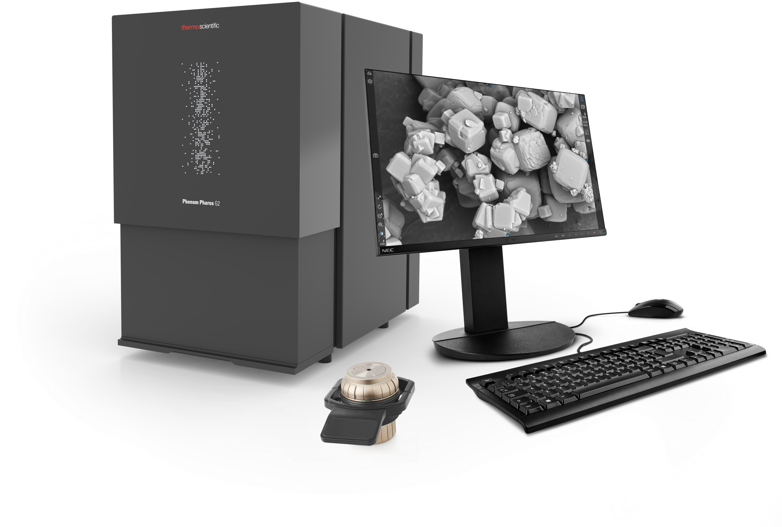
The Phenom Pharos scanning electron microscope is the latest model of microscope manufactured by Thermo Fisher Scientific. It is the first teenage microscope equipped with a FEG field-emission source, which not inconsiderably improves the sharpness and brightness of SEM images and provides a number of other advantages of using a FEG source. The microscope, like the other Phenom microscopes, is easy to use thanks to its compact design – starting with the installation process as well as the actual use. The advanced design of the device’s components and detectors means that electron images are obtained after tens of seconds, and the imaging process itself, despite the sophistication of the process, is simple and intuitive.
The Phenom Pharos scanning electron microscope is designed to quickly and extremely simply collect the highest quality electron images with magnifications of up to 2,000,000x and a real resolution of less than 2nm. This microscope, from the moment the sample is placed in the microscope chamber to the first electron image – takes only 30 seconds.
In addition, as with the other Phenom microscopes, we can use a number of sample holders with automatic tilt and rotation or temperature control, among others. The equipment can also be supplemented with application software for 3D reconstruction and automatic particle or porosity analysis, among others.
Using basic functions such as automatic focusing or astigmatism correction, the Phenom Pharos microscope offers to take high-quality electron images in the shortest possible time.
The user-friendly control software allows expansion with a range of additional image analysis programs.
Specification
Easy to use, intuitive user interface
Time required for image acquisition:
Optical: 5 seconds
Electron: 30 seconds
Magnification up to 2,000,000x
Extremely easy sample navigation to the point of interest
FEG electron source optimized for high-resolution capability
Comprehensive self-diagnostic system
With special sample holder – no risk of damage to electron optics components is achieved
Color navigation camera: perfect correlation between optical and electron-optical images
Backscattered electron detector, with the ability to operate in two modes:
Basic
Topographic
Ability to archive images in the following formats:
TIFF
JPEG
BMP
Possibility of collecting optical images
Basic system – great value for money
Very low power consumption
Resolution capability: < 2nm
Accelerating voltage: 1kV – 20kV
The microscope is equipped with a CCD camera for viewing and positioning the sample in the microscope.
Accessories
- Secondary electron detector (SED)
- EDS spectrometer (SDD type)
- Temperature-controlled holder
- Motorized eucentric holder with sample tilt and rotation
- Metallographic specimen holders (diameter up to 32mm)
- Software for automatic fiber recognition and analysis
- Software for automatic particle recognition and analysis
- Software for automatic pore recognition and analysis
- Software for 3D reconstruction of the sample surface
- Software for mapping and linear EDS analysis
- Libraries of microscope control functions, for use in .NET and Python environments