

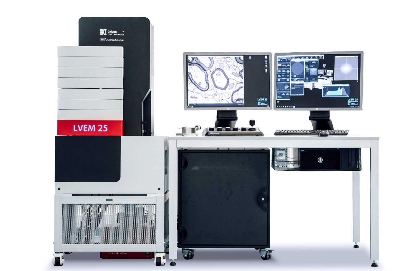
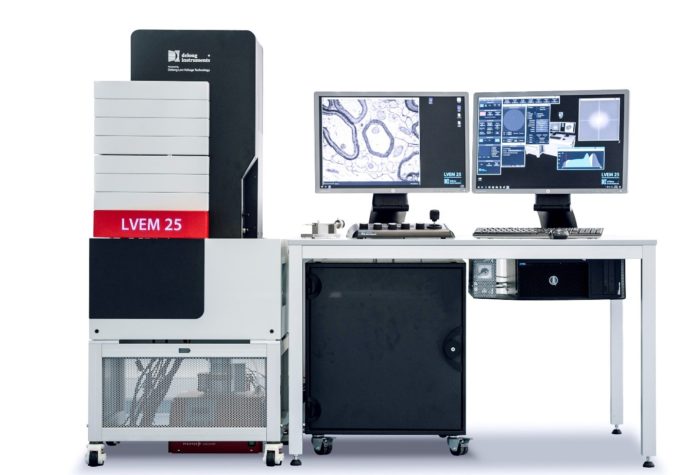
Transmission electron microscope LVEM25

Description
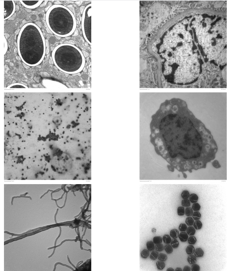
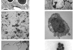
The LVEM 25 is a unique investigative tool which combines transmission TEM and STEM observation modes. Substantially lower accelerating voltages (ranging from 25 kV to 10 kV) than conventional TEM (typically 80–200 kV) provide substantially improved contrast on light elements with conventionally prepared samples. Low voltages result in increased electron scattering and enhanced contrast on biological, organic and light materials, without the need for staining.
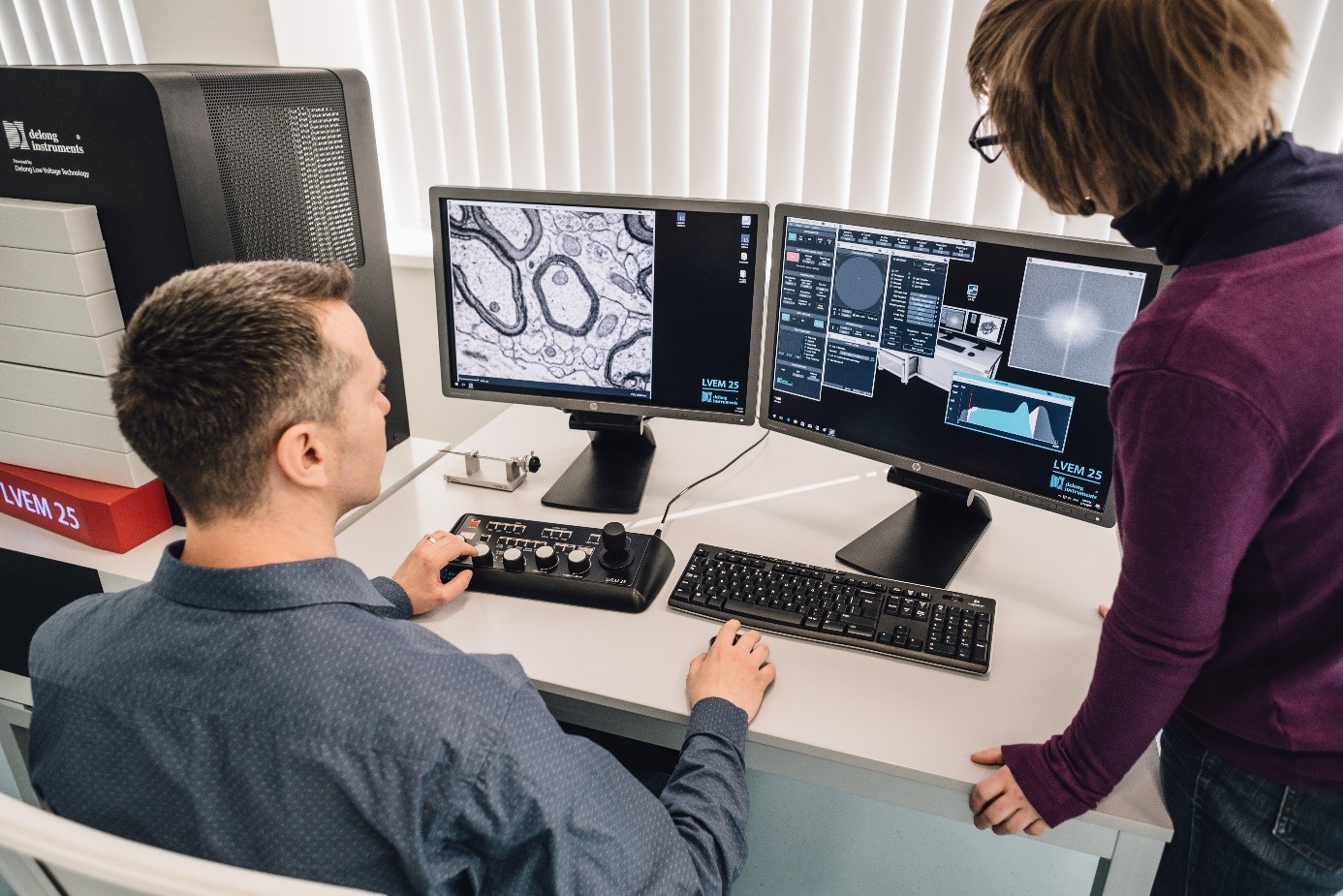
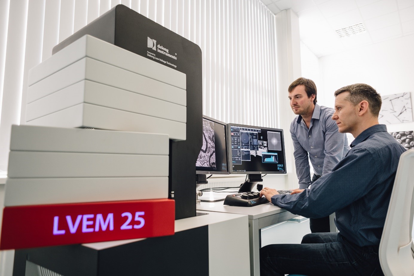
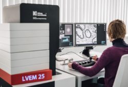
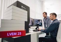
The LVEM 25 has an architecture that differs from traditional models. It can be installed in a lab, on a desktop or benchtop; almost anywhere electron imaging is needed. The system can even be supplied as a mobile work-station. The system has no special facilities requirements, no need for a dark room, cooling water, or special power. Ownership and maintenance of this system are greatly simplified.
- Powerful transmission electron microscope.
- The LVEM 25 combines transmission TEM and STEM modes.
- It operates with low accelerating voltage (25 kV) which brings enhanced contrast on light elements.
- Resolving power down to 1 nm.
- Biological and soft matter applications.
- High contrast.
- Imaging possible both with and without staining.
- Easy to control.
- Small installation space.
- The LVEM 25 can be installed in an ordinary lab without any need for a dark room or cooling water. It can be supplied even as a mobile work-station.
- Specimen tilt holder availability.
Specification
- electrons source with Schottky cathode (S-FEG) with a lifetime of> 2,000 hours;
- accelerating voltage 6, 10, 25 kV;
- time from sample insertion until the first electron photo is obtained ≤ 3 minutes;
- in the TEM imaging mode:
- resolving power ≤ 1 nm;
- maximum magnification ≥ 430,000x;
- saved images with a resolution of up to 2,048 x 2,048 pixels;
- in the STEM imaging mode:
- resolving power ≤ 1 nm;
- maximum magnification ≥ 470,000x;
- saved images with a resolution of up to 2,048 x 2,048 pixels;
- in ED diffraction mode:
- minimum beam diameter ≤500nm
- camera length 2000 – 7000 pixels
Consumables
https://micro-shop.pl/page/6/?s=siatki+TEM&post_type=product
For more supplies, please visit our online store Micro-Shop.