

Ultra-fast automated multibeam electron microscope

Description
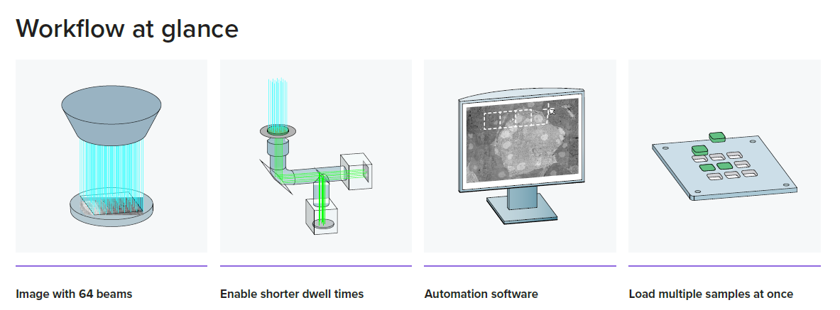
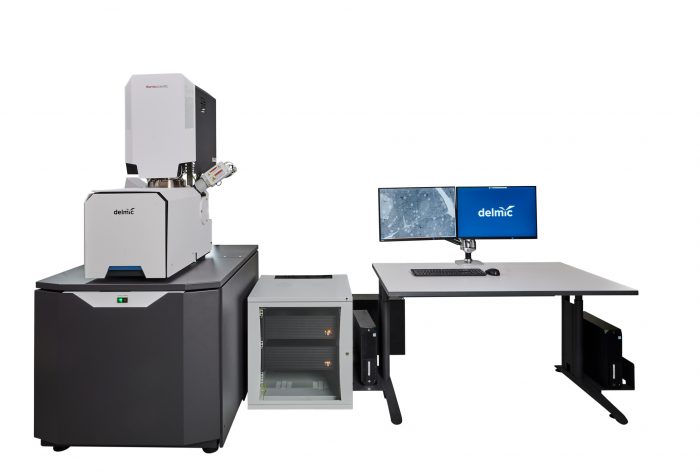
FAST-EM is an ultra-fast automated multibeam electron microscope (EM) designed to make complex and large EM projects simple and efficient. Thanks to its automated acquisition, this high-throughput system is ideal for imaging large or multiple samples for quantitative analysis. Delivering powerful insights while keeping the workflows simple, this system allows users to shift their focus from microscope operation to data analysis.
FAST-EM can be used to explore cell architecture, the interaction of neuronal circuits, and the analysis of any biological material. It is extremely beneficial for large volume 3D imaging, large scale 2D imaging and, in general, as a tool that can significantly speed up daily microscopy work.
Specification
| Electron optics | |||||
| System base | Thermo Fisher Scientific Apreo 2 | ||||
| Emitter: | Schottky field emission source | ||||
| Emitter stability: | Per 24 hours | 3% | |||
| High tension range: | 2.5-10 kV | ||||
| Beamlet current: | 400-800 pA | ||||
| Total current: | 25.6-51.2 nA | ||||
| Electron beam resolution: | (35-65% edge) | 4 nm | |||
| Nominal working distance: | 5 mm | ||||
| Single beam mode: | Yes | ||||
| Scanning and detection | |||||
| Multiprobe arrangement: | Square, 8 x 8 array | ||||
| Beamlets: | 64 | ||||
| Dwell time: | 400 ns minimum, adjustable | ||||
| Pixel size: | During field acquisition | 4 nm | |||
| Field of view: | At 3.2 μm pitch | 25.6 x 25.6 um | |||
| Detectors | Multibeam | Transmission detector with 64 silicon photomultiplier cells | |||
| Single-beam | Segmented in-lens backscattered electron detector | ||||
| Upper in-lens secondary electron detector | |||||
| Sample and stage | |||||
| Type | 3 axes motorized (XYZ) | ||||
| Stage position readout | Laser interferometry for nanometer-level positioning accuracy | ||||
| Travel range XY | 50 × 50 mm | ||||
| Typical substrate size | 14 x 14 mm* | ||||
| Max simultaneous substrates | When using 14 × 14 mm substrates | 9 – for a total of ~850 1*1 mm sections | |||
| Imaging workflow | |||||
| Usable samples | Directly on scintillators | Resin sections (maximum thickness of 150 nm), nanoparticles, vesicles, viruses | |||
| On TEM grids** | Resin sections (maximum thickness of 150 nm), nanoparticles, vesicles, viruses | ||||
| Unattended run-time | 72 hours | ||||
| Use cases | Routine data collection | Semi-automated imaging of user-defined ROIs and section arrays | |||
| Sustained throughput | During megafield acquisition at 400 ns | 100 megapixels/second | |||
| Data format | One 16-bit TIFF per field image, stored per project | ||||
| Software | |||||
| Microscope control | Linux-based acquisition control | ||||
| Acquisition support | User guidance for basic operations | ||||
| System health monitoring | Continuous logging of crucial system features | ||||
| Automatic calibrations | Detector gain, detector alignment, autostigmation, autofocus, global alignment of components | ||||
| Vacuum and support hardware | |||||
| Vacuum pumps | Turbomolecular pump, scroll pump (backing) | ||||
| Operational vacuum | ≤ 3 × 10⁻⁵ mbar | ||||
| Network storage connection | 10 Gbit Ethernet (10GBASE-SR using LC Duplex OM3 MM fiber) | ||||
| Optional components | |||||
| High performance storage module | Scalable high-speed storage for data analysis and data sharing | ||||
| Support Infrastructure | Standalone water chiller | ||||
| Acoustic enclosure for backing pump | |||||
| Consumables | 14 × 14 × 0.15 mm scintillator substrates | ||||