

Fluorescence light microscope (FLM) integrated into a FIB/SEM system
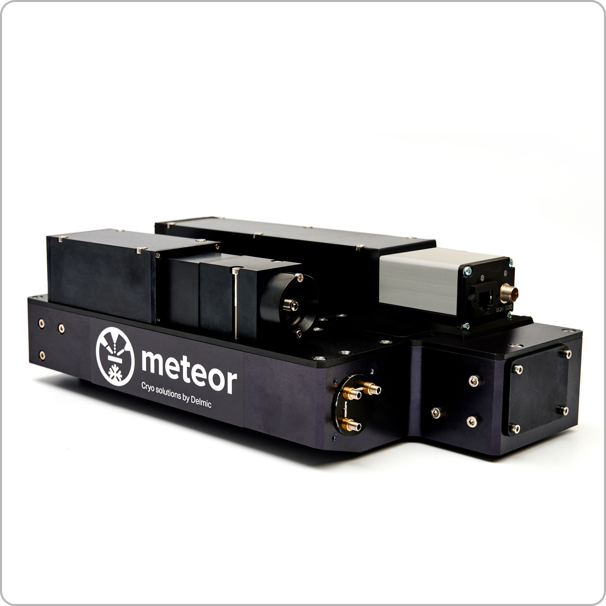
Description
METEOR overcomes the challenges in the current cryogenic electron tomography (cryo-ET) workflow by providing the capability to perform in situ fluorescence light microscopy (FLM) in your cryo focused ion beam (FIB)/ scanning electron microscope (SEM) chamber. Not only does it reduce the number of transfer steps between microscopes, protecting the fragile sample from unnecessary contamination, it will also allow you to confirm the presence of the region of interest (ROI) in the lamella directly after FIB milling. METEOR is highly adaptable to your current workflow and works well with your transfer systems and sample holders, making it easy to adopt.
METEOR can ensure an efficient workflow and increased sample yield for downstream acquisition of high resolution three dimensional biological structures in their near-native cellular environment. It’s a powerful tool that can be applied to a wide range of research areas including microbiology, neurosciences, immunology, virology, developmental, cell and molecular biology.
Increase CRYO-ET sample yield
Target your ROIs and correlate better and more effectively with the integrated FLM. Reduce transfer steps and thereby sample damage.
Optimize your CLEM workflow
Save time and work more efficiently by having both FLM and FIB in the same microscope
Boost productivity
Produce high quality lamellae more easily. Obtain insight into your biological system through getting useful tomography data more quickly.
Improve cost efficiencies
Use your cryo TEM time more effectively on useful lamellae. Remove the need for a separate cryo FLM dedicated solely to ROI finding.
Specification
System specifications
Compatible FIB/SEM systems
- Thermo Fisher Scientific Aquilos
- Thermo Fisher Scientific Helios
- Thermo Fisher Scientific Scios
- Zeiss Crossbeam 550
- Zeiss Crossbeam 550L
Optics
Objectives: the following options are available*
- Olympus Fluorite objective:
○ 10x (WD 11.0 mm, NA 0.30)
○ 20x (WD 3.1 mm, NA 0.45)
○ 50x (WD 1.0 mm, NA 0.80)
○ 100x (WD 1.0 mm, NA 0.90)
- Olympus Apochromat objective:
○ 50x (WD 0.35 mm, NA 0.95)
○ 100x (WD 0.35 mm, NA 0.95)
- Olympus Semi-Apochromat objective:
○ 50x (WD 10.6 mm, NA 0.50)
* Depending on shuttle and sample stage configuration the choice of
objectives can be limited due to space restrictions
Objectives stage:
- Travel range: 31 mm
- Minimal incremental motion < 50 nm
Lightsource:
Omicron LedHub fitted with 4 LED sources:
- 385 nm
- 470 nm
- 505 – 600 nm – bandpass filter
- 625 nm
Camera: the following options are available
- sCMOS camera – 6.5 μm pixel size (Andor Zyla 4.2)
- sCMOS camera – 6.5 μm pixel size (Andor Sona 4.2B-6)
Filters:
Equipped with a filter wheel with four slots, several multiband or single-band configurations are possible that can be optimized to the user’s needs. Example configuration:
- 440/40 nm single-band bandpass filter
- 525/30 nm single-band bandpass filter
- 607/36 nm single-band bandpass filter
- 684/24 nm single-band bandpass filter
Imaging modes:
- Fluorescence imaging
- Reflection imaging
Dimensions
The METEOR system consists of 3 main parts. The electronics rack (placed in the microscope room), the body (mounted on the FIB/SEM) and the in chamber optics (mounted inside the FIB/SEM). The dimensions (H/W/D) are given below. Please note that depending on the exact configuration chosen these dimensions might change.
- Electronics rack (80 cm x 60 cm x 80 cm)
- METEOR body (30 cm x 30 cm x 20 cm)
- METEOR in-chamber ( 6 cm x 6 cm x 20 cm)
System Control
Comes with a control PC next to the FIB/SEM computer for acquiring your fluorescence images.
Odemis Integrated Software
ODEMIS, a user-friendly open-source acquisition software, is installed together with METEOR. It will ensure image acquisition parameters are primed for high quality fluorescence images. Users can furthermore implement their own scripts in Python to automate routine processes, e.g. camera exposure time optimisation.
Software functions:
- Adjust all relevant imaging parameters (LED power, camera exposure time and gain, excitation and emission filter)
- Tiling and stitching functionality to acquire large sample areas overview
- Control of the FIB/SEM stage to navigate the sample and find the region of interest
- Switch between the SEM and FLM positions to acquire new FLM images as materials are milled away
- Save sample and objective stage coordinates to quickly reinspect milled lamellae
- Switch between SEM and FLM positions to verify fluorescence signals in the lamella
- Z-stack acquisition
- Camera pixel binning option
- Multicolour imaging
- Odemis viewer to inspect the images
- Licence-free (free updates)