

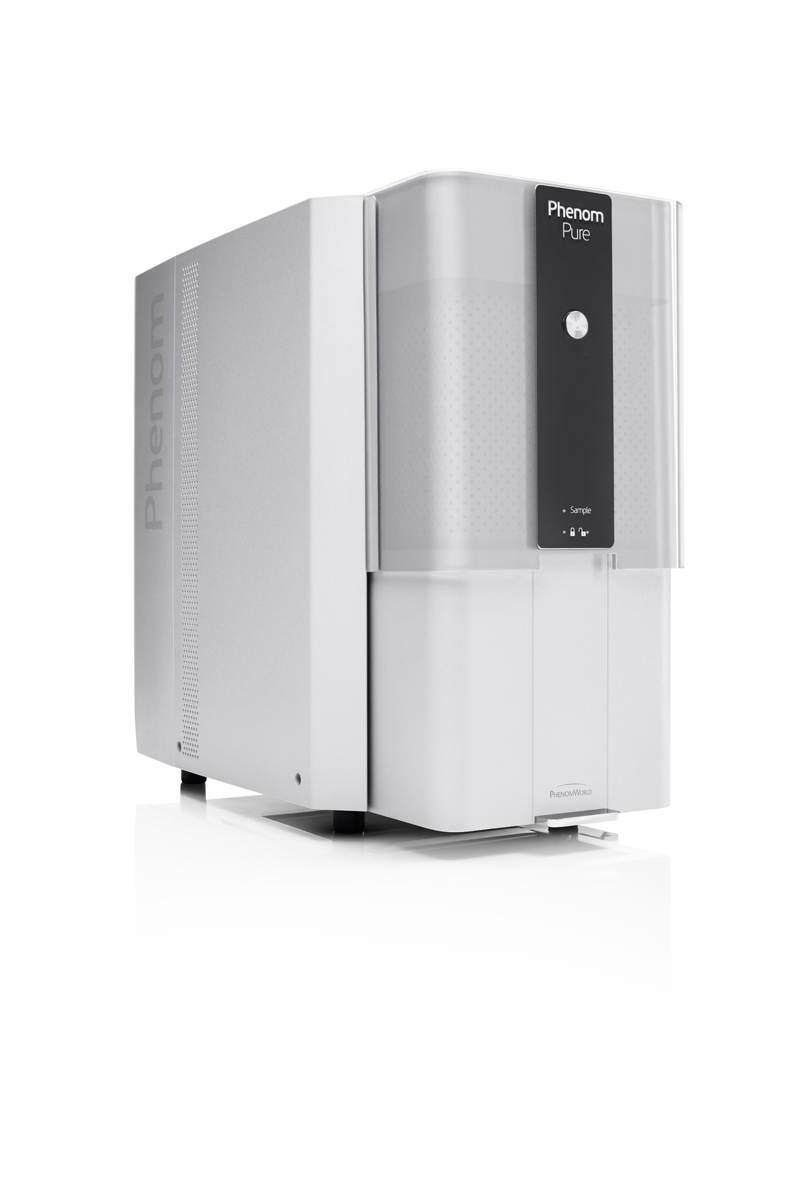
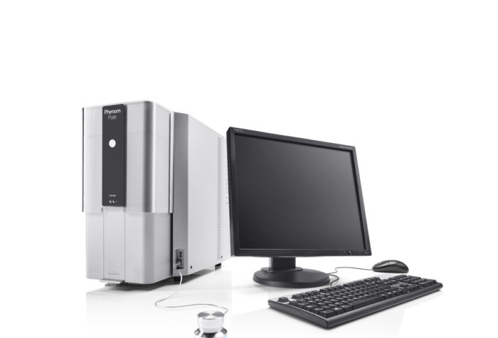
Phenom Pure

Description
The Phenom Pure desktop scanning electron microscope (SEM) is an ideal tool for making the transition from working with a light microscope to operating on electron microscope. It is equipped with the basic fundamentals for meeting imaging needs.
The Phenom Pure provides high-quality images using basic features, and offers the market’s fastest loading and imaging time. The reliable autofocus, automatic stigmator and source alignment make this the most user-friendly system on the market and enable acquiring images of best quality with the shortest acqusition time on the market. It is the most economic and efficient tool for high-resolution imaging.
The Phenom Pure microscope is the fastest SEM in the market. The time from loading the sample up to acquiring SEM image is less than 30 seconds.
The Phenom Pure is equipped with highly efficient CeB6 (Cerium Hexaboride) electron source which is characterized by high signal to noise ratio and high image resolution. User-friendly software can be extended by many additional features like automatic image analysis.
Its worry-free maintenance is unique in its product category and maximizes system uptime. With these characteristics, the Phenom Pure can be operated by any staff member, bringing high-magnification imaging within the reach of all lab personnel.
The Phenom Pure is a complete and ready-to-go system. Unpack, install and it is ready for action without the need for a PC, laptop or other peripherals. The Phenom Pure is the most stable SEM microscope on the market. There is no need of using any antivibration system.
The Phenom Pure microscope can be easly upgraded up to Phenom ProX model
Specification
- Easy to operate, intuitive user interface
- Sample loading time:
• Light optical: 5 seconds
• Electron optical: 30 seconds - Magnification range: up to 175 000X
- Image resolution < 15 nm
- Easy sample navigation
- Long lifetime electron source (CeB6)
- Optimized electron source for obtaining high imaging resolution
- Versatile autodiagnostic system
- Unique sample holders eliminating any risk of damage of detectors o rany part of vacuum system
- Acceleration voltage: 5kV – 10kV
- Monochromatic CCD navigation camera: perfect correlation between light optical and electron optical images
- Back scattered electrons detector with two modes of imaging:
• Full
• Topographic - Images formats:
• TIFF
• JPEG
• BMP - Excellent quality to price ratio
- Low power consumption
Accesories
- Secondary electron detector (SED)
- Temperature controlled sample holder
- Motorized eucentric sample holder (tilt and rotation)
- Resin mount sample holder (diameter up to 32mm)
- Software for automatic fibers recognition and measurement
- Software for automatic particles recognition and measurement
- Software for automatic pores recognition and measurement
- Software for 3D roughness reconstruction
- Software for EDS mapping and linescan
Consumables
10-002012-100 – Pin stubs for SEM, Ø12.7, standard pin, aluminium (Pack of 100).
10-002025-10 – Pin stubs for SEM, Ø25.4, standard stub, aluminium (Pack of 100).
AGG3347N – Adhesive carbon tabs, Agar Scientific 12 mm (Pack of 100)
AGG3348N – Adhesive carbon tabs, Agar Scientific 25 mm (Pack of 50)