

SECOM-Integrated Correlative Light and Electron Microscope
Description
The SECOM platform is a unique integrated solution for correlative light and electron microscopy that enables you to do correlative microscopy extremely fast with the highest optical quality and overlay accuracy. The combination of the labeling power of fluorescence imaging and the high resolution structural information of electron microscopy makes correlative microscopy the perfect tool to study the complex relation between form and function in biology.
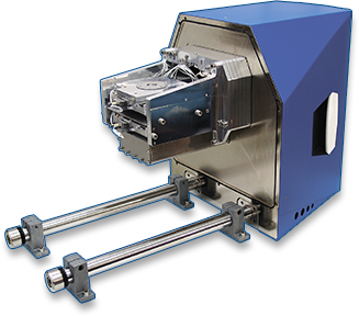
The SECOM is the only product to date that integrates fluorescence and scanning electron microscopy in one device by equipping a scanning electron microscope (SEM) with an inverted fluorescence microscope. The platform can be fitted on a SEM by replacing the door to the chamber of the SEM. This replacement supports a motorized stage and the objective and light path for the optical microscope.
Thanks to its integrated design, switching from fluorescence to electron imaging is seamless and instantaneous. And with the automated alignment procedure, you are directly imaging the right location at a high resolution. The SECOM platform comes with an intuitive software package that is designed to easily acquire both types of information. The software package can also control the SECOM stage and the settings for the electron microscope.
The SECOM platform can easily be integrated in the existing workflow and acts like a fully equipped high-end optical microscope, without compromise on either optical or electron performance.
Specification
Excitation
- Multiband setup in Pinkel configuration optimized for DAPI, FITC, TRITC, & Cy5 and other like fluorophores.
- Four channel solid-state (LED) excitation source with digital On/Off and intensity control.
- Default excitation wavelengths at 387/11, 485/20, 560/25 and 650/13nm.
Others available on request.
Objective Stage
- Stage uses piezoelectric stepping motors that remain full blocking force when not actuated, resulting in small thermal load and thus low drift.
- Minimum incremental motion in XY less than 500 nm.
- Repeatability of Z-axis (focusing) 50 nm.
- Use of optical linear encoder for closed loop driving.
Sample Stage
- Total stroke of 18x18mm in XY.
- Equipped with precision piezoelectric stepping motors and optical linear encoders.
- Minimum incremental motion of 300 nm, repeatability of 500 nm.
Camera
- Scientific CMOS camera allows imaging with low noise and large field of view.
- 2048×2048 pixels with a pixel size of 6.5 μm yields
- 330×330 μm field of view at 40x magnification
Objective lens
- Plan Apochromat objective lens, magnification 40x, numerical aperture 0.95.
- Other objective lenses, including immersion lenses (up to NA 1.4), available on request.
- User exchangeable