Phenom ParticleX TC
Decription
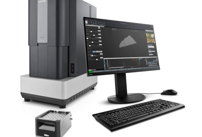
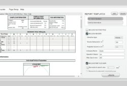
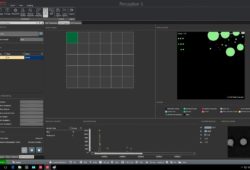
The Phenom Particle X Technical Cleanliness Scanning Electron Microscope is a multipurpose SEM enabling automated cleanliness analysis according to VDA 19.1 and ISO 16232. This microscope is based on Phenom XL model. ParticleX is able to measure particles on pre-prepared filters from 500 nm to 3mm. Our system is equipped with EDS spectrometer which allows us to collect compositional data from all particles on filter. Particle X is an invaluable tool for examination extracted particles and tracking the source of particle contamination during production, transportation or packing. As a standard Particle X has integrated reporting software fully compliant with VDA 19.1 and ISO 16232. This is world’s first desktop SEM that can run fully automated Cleanliness analysis with four standardized filters 47 mm. Both software and hardware are fully integrated to enhance user-friendliness, reliability and analysis speed.
The Phenom ParticleX TC Electron Microscope (SEM) pushes the boundaries of compact desktop SEM performance. It features the proven ease-of-use and fast time-to-image of any Phenom system.
Like in case of other Phenom microscopes this model is characterized by user friendly operation software.
It is also equipped with a chamber that allows analysis of large samples up to 100 mm x 100 mm (65mm of height). A proprietary venting/loading mechanism ensures the fastest vent/load cycle in the world, providing the highest throughput. A newly developed compact motorized stage enables the user to scan the full sample area, and yet the Phenom ParticleX is a desktop SEM that needs little space and no extra facilities.
Ease-of-use is given an extra boost in the Phenom ParticleX with a single-shot optical navigation camera that allows the user to move to any spot on the sample with just a single click – within seconds.
With its long-life high-brightness CeB6 electron source, the Phenom ParticleX creates state-of-the-art images with a minimum of user maintenance intervention.
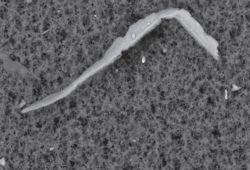
The ParticleX microscope is dedicated to easly acquire SEM images of highest quality with magnification up to 100 000x and image resolution less than 14nm.
The ParticleX microscope is the one of the fastest SEM in the market. The time from loading the sample up to acquiring SEM image is less than 60 seconds.
A large variety of sample holders are available for facilitating the fast loading of any sample into the Phenom.
The ParticleX is equipped with highly efficient CeB6 (Cerium Hexaboride) electron source which is characterized by high signal to noise ratio and high image resolution.
The backscattered detector (BSD) and detection chain are optimized to work together and give results with unmatched signal-to-noise images on a large variety of samples.
The Phenom ParticleX can be upgraded with the ProSuite application platform. ProSuite offers a variety of software applications that will automate data collection and image interpretation.
Its worry-free maintenance is unique in its product category and maximizes system uptime. With these characteristics, the Phenom ParticleX can be operated by any staff member, bringing high-magnification imaging within the reach of all lab personnel.
The Phenom ParticleX is a complete and ready-to-go system. Unpack, install and it is ready for action without the need for a PC, laptop or other peripherals.
Specification
- Easy to operate, intuitive user interface fully compliant with VDA 19.1 and ISO 16232.
- Sample loading time:
- Light optical: 5 seconds
- Electron optical: 60 seconds
- Magnification range: 80 – 100 000X.
- Image resolution <14nm
- EDS spectrometer to collect compositional data
- Easy sample navigation.
- Long lifetime electron source (CeB6)
- Optimized electron source for obtaining high imaging resolution.
- Versatile autodiagnostic system.
- Unique sample holders eliminating any risk of damage of detectors o rany part of vacuum system.
- Acceleration voltage: 5kV – 20kV.
- CCD navigation camera: perfect correlation between light optical and electron optical images.
- Images formats:
- TIFF
- JPEG
- BMP
Excellent quality to price ratio.
Low power consumption.
- Sample loading time:
Accesories
- Secondary electron detector (SED)
- Software for automatic pores recognition and measurement
- Software for automatic fibers recognition and measurement
- Software for 3D roughness reconstruction
- Software for EDS mapping and linescan
Consumables
https://micro-shop.pl/produkt/peseta-wafli-typ-34a/
https://micro-shop.pl/produkt/tasma-weglowa/
https://micro-shop.pl/produkt/krazki-weglowe/
For more supplies, please visit our online store Micro-Shop.


